Back

Role of tangential spot-compression view in assessment of a small palpable mass turned out to be a Multi-focal disease

Author / Copyright: Dr. Eman Ewais (Breast Imaging Specialist and Educator)

Clinical History

A 50 yrs old generally healthy female patient presented by a recently felt palpable lump in her left breast she has no personal history of breast cancer or breast surgery. Positive family history of breast cancer (sister at post-menopausal age).

Case Imaging Scenario

Patient was initially assessed by digital mammography standard views (bilateral mediolateral oblique (MLO) and craniocaudal (CC) views of the breasts), which showed a small focal asymmetry at the LOQ middle third of the left breast about 4 o’clock in position. [Figure 1]. Right breast mammogram was normal.

Targeted evaluation by hand held high resolution ultrasound was performed and showed a small papion-shaped, irregular, taller than wider, hypoechoic solid lesion at the left breast 4 o’clock corresponding to the mammographic focal asymmetry location and patient area of complaint. It measured about 8 x 6 x 5 mm in maximum dimensions. Doppler ultrasound showed an abnormal peripheral vessel. It was also surrounded by an echogenic halo. Findings were of high suspicion of malignancy. [Figure 2].

The initially noted mammographic findings were poorly characterized as a mass despite the low density of this ACR B breast, so, there was a need to confirm the concordance between mammography and ultrasound findings and It was notable in ultrasound that the lesion is superficial in location so an additional tangential spot-compression view was performed at 60o angle and showed an irregular bi-lobed mass of high density and irregular margin consistent with ultrasound suspicious findings [Figure 3].  The mass was given a BI-RADS category 4c and referred for biopsy which confirmed a diagnosis of invasive ductal carcinoma.

Palpable breast lumps has a high yield of breast cancer with PPV up to 80%  [1], yet it is not usually felt when it is less than 1 cm unless it is superficial in location which is unusual for breast cancer. Unless suspicious imaging features is seen, it could be mistaken for other breast lesions like dermal and subcutaneous fat lesions [2].

Mammography can be misleading in some cases, due to overlapping of glandular densities or technical insufficiency of the standard views for certain anatomic location of some lesions. Yet, additional views like spot compression, magnification and tangential views can allow correct prediction of benign and malignant breast lesions by improving the visualization and characterization of lesions at the examined region of interest [2] [3].

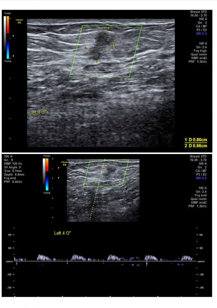
In this Case, assessment by mammography was the initial modality of choice as the patient is above the age of 40 and she is presenting with a palpable lump, yet mammography only showed a focal asymmetry that was also not calcified and no associated architectural distortion which lowered the prediction of malignancy. But the ultrasound findings were more suspicious, despite the superficial location of the lesion which could be mistaken for an IMLN considering the initial mammographic findings.  So, an additional mammographic view was needed to confirm help the radiologist in reaching an appropriate BI-RADS category.

When the additional tangential spot-compression view was performed, It was very clear the similarity between the visualized mass and the ultrasound findings as bi-lobed irregular mass. No lucent halo or fat density was identified within the mass; rising the suspicion of the lesion.

A final assessment of BI-RADS category 4c was given and biopsy was recommended.

  1. Teaching Points:

    • Tangential views can help in visualization of superficial breast lesions if obscured in standard views.
    • Special mammographic views can play an important role in characterization of palpable breast lumps.
    • Mammographic views can be adjusted to match the diagnostic needs and the radiologist can use them to be more confidence in assigning the final BI-RADS category.


    Final Diagnosis:                                                                                           

    Multi-focal Invasive ductal carcinoma GII; two small masses 2 mm apart.
    ER –ve, PR –ve, Her2/neu strong +ve .

    Negative sentinel lymph node.


    Differential Diagnosis List:

    • Ductal carcinoma.
    • Lobular carcinoma
    • IMLNs
    • Fat necrosis.

    Keywords: Additional views, spot compression, multi-focal IDC, Her2 positive, tangential view, breast cancer, early detection

  1. Karim M, Khan K A, Khan A, et al. (March 30, 2020) Triple Assessment of Breast Lump: Should We Perform Core Biopsy for Every Patient?. Cureus 12(3): e7479. DOI 10.7759/cureus.7479.
  2. Giess CS, Raza S, Birdwell RL. Distinguishing breast skin lesions from superficial breast parenchymal lesions: diagnostic criteria, imaging characteristics, and pitfalls. Radiographics. 2011;31(7):1959-1972. doi:10.1148/rg.317115116
  3. Faulk R M, Sickles E A. Efficacy of spot compression-magnification and tangential views in mammographic evaluation of palpable breast masses. Radiology 1992 185:1, 87-90.
  1. All submitted images of this case was performed at techno scan women’s health canter, Tagmoa branch, Cairo, Egypt.

Leave A Reply